
Nocturnal Ventilatory Polygraphy: Prescription, Mechanism & Interpretation
The essential home sleep test for diagnosing sleep apnea and hypopnea without hospitalization. Discover how this ambulatory examination works, what the Apnea-Hypopnea Index (AHI) reveals about your breathing, and when polysomnography may be required for a complete diagnosis.
What Is Nocturnal Ventilatory Polygraphy?
Nocturnal ventilatory polygraphy (also called respiratory polygraphy or home sleep apnea test) is a simplified diagnostic examination designed to detect and quantify sleep-disordered breathing—particularly obstructive sleep apnea syndrome (OSA). Unlike full polysomnography which requires hospitalization, this ambulatory test is performed at home, making it more accessible, convenient, and cost-effective for millions of patients worldwide.

The examination monitors several physiological parameters during sleep—including airflow, breathing effort, blood oxygen saturation, and heart rate—to identify apneas (complete breathing pauses) and hypopneas (partial airflow reductions). These measurements generate the critical Apnea-Hypopnea Index (AHI), the primary metric used to diagnose and classify the severity of sleep apnea.
💡 Why This Test Matters
Sleep apnea affects an estimated 936 million adults globally—yet up to 80% remain undiagnosed. Untreated OSA significantly increases the risk of cardiovascular disease, stroke, hypertension, type 2 diabetes, and even premature death (up to 46% higher mortality risk according to Johns Hopkins research). Nocturnal ventilatory polygraphy provides the accessible diagnostic pathway needed to identify and treat this condition before serious complications develop.
Who Should Undergo Nocturnal Ventilatory Polygraphy?
This diagnostic examination is prescribed for patients presenting symptoms suggestive of sleep-disordered breathing. Your physician may recommend respiratory polygraphy if you experience any of the following indicators:
Chronic Snoring
Ronchopathy (habitual snoring) affecting sleep quality for you or your partner. Snoring occurs in 44% of men and 28% of women.
Witnessed Apneas
Breathing pauses observed by a bed partner, often followed by gasping, choking, or loud snorts as breathing resumes.
Excessive Daytime Sleepiness
Persistent fatigue, difficulty concentrating, morning headaches, or falling asleep during daily activities despite adequate sleep time.
Cardiovascular Risk Factors
Resistant hypertension, atrial fibrillation, heart failure, stroke history, or type 2 diabetes—conditions strongly linked to OSA.
Clinical Indications for Respiratory Polygraphy
| Symptom Category | Specific Indicators | Risk Level |
|---|---|---|
| Nighttime Symptoms | Loud snoring, witnessed apneas, gasping/choking, restless sleep, nocturia (frequent urination), night sweats | High suspicion |
| Daytime Symptoms | Excessive sleepiness, morning headaches, dry mouth/sore throat upon waking, irritability, memory/concentration problems | High suspicion |
| Physical Characteristics | Obesity (BMI >30), large neck circumference (>43cm men, >38cm women), retrognathia, enlarged tonsils | Increased risk |
| Comorbidities | Resistant hypertension, atrial fibrillation, heart failure, stroke, type 2 diabetes, pulmonary hypertension | Screening recommended |
⚠️ When Polygraphy Is NOT Sufficient
Respiratory polygraphy is designed specifically for suspected obstructive sleep apnea. Patients with the following conditions require full polysomnography instead:
- Suspected central sleep apnea or complex sleep disorders
- Significant cardiopulmonary disease (COPD, heart failure, neuromuscular disorders)
- Suspected parasomnias (sleepwalking, REM behavior disorder)
- Suspected periodic limb movement disorder or restless legs syndrome
- Children under 18 (pediatric protocols differ significantly)
- Patients requiring supplemental oxygen or mechanical ventilation
How Nocturnal Ventilatory Polygraphy Works: The Mechanism
Respiratory polygraphy employs a portable monitoring device equipped with multiple sensors that record breathing patterns and physiological responses throughout the night. Unlike polysomnography, it does not monitor brain activity (EEG), making it simpler to administer at home without technical supervision.

Sensor Placement & Measurements
Several sensors are strategically positioned on the patient's body to capture comprehensive respiratory data:
🫁 Nasal Cannula
Location: Nose
Measures: Oronasal airflow through pressure changes, detecting apneas and hypopneas in breathing
💨 Oral Thermistor
Location: Near mouth
Measures: Oral airflow via temperature changes, capturing mouth breathing patterns
📊 Thoracic Band
Location: Chest
Measures: Thoracic respiratory movements using inductance plethysmography (RIP)
📈 Abdominal Band
Location: Abdomen
Measures: Abdominal respiratory effort, distinguishing obstructive from central apneas
❤️ Pulse Oximeter
Location: Finger (typically index or ring)
Measures: Oxygen saturation (SpO2) and heart rate throughout the night
🎤 Snoring Sensor
Location: Neck or integrated
Measures: Snoring intensity and frequency via vibration detection
🧭 Position Sensor
Location: Chest or device
Measures: Body position (supine, lateral, prone) to correlate with respiratory events
💓 ECG (Optional)
Location: Chest electrodes
Measures: Heart rhythm and rate, detecting cardiac arrhythmias associated with apneas
What the Test Records
| Parameter | What It Measures | Clinical Significance |
|---|---|---|
| Airflow (nasal pressure) | Breathing amplitude and frequency | Identifies apneas (≥90% reduction) and hypopneas (≥30% reduction) |
| Respiratory Effort | Thoraco-abdominal movements | Distinguishes obstructive (effort present) from central (no effort) apneas |
| SpO2 (Oxygen Saturation) | Blood oxygen levels | Quantifies desaturation events; severity indicated by drops below 90% |
| Heart Rate | Pulse frequency | Detects bradycardia/tachycardia associated with apneic events |
| Body Position | Supine, lateral, prone | Identifies positional sleep apnea (worse when sleeping on back) |
| Snoring | Sound/vibration intensity | Correlates with airway obstruction; quantifies ronchopathy severity |
🔬 The Science Behind Respiratory Events
Apnea occurs when airflow through the nose and mouth stops completely (≥90% reduction) for at least 10 seconds. This triggers oxygen desaturation and often micro-arousals as the brain signals the body to resume breathing.
Hypopnea is a partial airflow reduction (≥30%) lasting at least 10 seconds, accompanied by either ≥3% oxygen desaturation or an arousal. Both events contribute to the AHI calculation.
The Procedure: Step-by-Step Guide
Nocturnal ventilatory polygraphy is designed for simplicity and patient comfort. Here's exactly what to expect before, during, and after your home sleep test:
Before the Test: Preparation
Medical Consultation
Your physician evaluates symptoms and prescribes the test
Device Collection
Pick up the portable polygraph from clinic or receive by mail
Instructions Review
Healthcare technician explains sensor placement and device operation
Day-of Preparation
Avoid alcohol, sedatives, and caffeine; follow normal sleep routine
During the Test: Night of Recording
🌙 What Happens During Your Sleep Study
- Apply sensors before bedtime: Position the nasal cannula, finger oximeter, chest and abdominal bands, and any additional sensors as instructed
- Activate the device: Press the start button to begin recording (some devices start automatically)
- Sleep normally: Go to bed at your usual time; try to sleep in your typical position
- Recording duration: The device records for 6-8 hours minimum (aim for your full sleep period)
- Morning removal: Remove sensors upon waking and turn off the device
After the Test: Analysis & Results
Device Return
Return the equipment to the clinic or mail it back as instructed (usually within 24-48 hours)
Data Download
Sleep technicians transfer recorded data to specialized analysis software
Manual Scoring
Expert sleep physicians analyze the tracing, identifying and classifying each respiratory event
Report Generation
Comprehensive report generated including AHI, oxygen desaturation index, and other metrics
Results Consultation
Follow-up appointment with your physician to discuss findings and treatment recommendations
📋 Preparation Tips for Accurate Results
- Avoid alcohol for at least 3-4 hours before bedtime (relaxes throat muscles, may worsen apnea artificially)
- Skip sedatives/sleeping pills unless prescribed and discussed with your doctor
- Maintain your usual sleep schedule—don't try to "sleep better" for the test
- Sleep in your normal position (the test should capture your typical breathing patterns)
- Ensure secure sensor placement—loose sensors can cause signal loss and require repeat testing
- Record your bedtime and wake time to help technicians calculate accurate sleep time
Interpreting Your Results: Understanding the AHI Score
The Apnea-Hypopnea Index (AHI) is the cornerstone metric of respiratory polygraphy interpretation. It represents the average number of apneas and hypopneas per hour of sleep (or recording time for home tests), providing an objective measure of sleep-disordered breathing severity.

AHI Severity Classification (Adults)
| Severity Level | AHI Range (events/hour) | Clinical Interpretation | Typical Symptoms |
|---|---|---|---|
| Normal | < 5 | No significant sleep apnea | Occasional snoring possible; no treatment required |
| Mild OSA | 5 - 14 | Mild obstructive sleep apnea | Snoring, mild daytime fatigue; lifestyle changes often sufficient |
| Moderate OSA | 15 - 29 | Moderate obstructive sleep apnea | Significant sleepiness, witnessed apneas; treatment recommended |
| Severe OSA | ≥ 30 | Severe obstructive sleep apnea | Major symptoms, cardiovascular risk; treatment essential |
Pediatric AHI Interpretation
👶 Important: Children Use Different Thresholds
Because children have faster respiratory rates and smaller lung capacity, even fewer breathing events can be clinically significant:
- Normal: AHI < 1 event/hour
- Mild OSA: AHI 1 - 4.9 events/hour
- Moderate OSA: AHI 5 - 9.9 events/hour
- Severe OSA: AHI ≥ 10 events/hour
An AHI ≥ 1 in children is considered abnormal and warrants further evaluation.
Additional Metrics in Your Report
| Metric | Definition | Normal Range | Clinical Significance |
|---|---|---|---|
| ODI (Oxygen Desaturation Index) | Number of ≥3% oxygen drops per hour | < 5 events/hour | Correlates with AHI; indicates hypoxemic burden |
| Mean SpO2 | Average oxygen saturation during sleep | ≥ 94% | Lower values indicate chronic hypoxemia |
| Minimum SpO2 | Lowest oxygen saturation recorded | ≥ 90% | Below 80% = severe desaturation |
| T90 (Time below 90%) | Percentage of time with SpO2 < 90% | < 1% | Higher values indicate significant nocturnal hypoxemia |
| Snoring Index | Percentage of sleep time with snoring | Variable | Quantifies ronchopathy severity |
| Supine AHI | AHI specifically in supine position | Compare to overall AHI | If significantly higher → positional sleep apnea |
📊 When AHI ≥ 30: Polygraphy Is Typically Sufficient
According to clinical guidelines, when respiratory polygraphy reveals an AHI ≥ 30 (severe OSA), the diagnosis is clear and treatment can be initiated without additional testing. However, if AHI is lower than expected given clinical symptoms, or if other sleep disorders are suspected, polysomnography may be recommended for comprehensive evaluation.
Polygraphy vs. Polysomnography: Understanding the Differences
While both tests diagnose sleep-disordered breathing, they differ significantly in complexity, setting, and diagnostic capabilities. Understanding these differences helps you know what to expect and when each test is appropriate.
| Feature | Respiratory Polygraphy | Polysomnography (PSG) |
|---|---|---|
| Setting | Home (ambulatory) | Sleep laboratory (hospitalization) |
| Supervision | Unattended/self-administered | Attended by sleep technicians |
| Channels Recorded | 4-7 channels (respiratory focus) | 16+ channels (comprehensive) |
| Brain Activity (EEG) | ❌ Not recorded | ✅ Full EEG monitoring |
| Sleep Staging | ❌ Cannot determine sleep stages | ✅ Full hypnogram (REM, NREM stages) |
| Eye Movements (EOG) | ❌ Not recorded | ✅ Monitored for REM detection |
| Leg Movements (EMG) | ❌ Not recorded | ✅ Detects periodic limb movements |
| Cost | Lower (typically 50% less) | Higher (facility + staffing costs) |
| Wait Time | Shorter (often immediate) | Longer (weeks to months) |
| Comfort | Sleep in own bed | Unfamiliar environment |
| Technical Failure Rate | 5-20% (may require repeat) | < 5% (technician intervention) |
| Primary Diagnosis | Obstructive sleep apnea | All sleep disorders |
When Polysomnography Is Required
🏥 Indications for Full Polysomnography
Despite polygraphy's convenience, certain clinical scenarios mandate comprehensive polysomnography:
- Negative polygraphy with high clinical suspicion: When symptoms strongly suggest OSA but polygraphy is negative or inconclusive
- Suspected central sleep apnea: Requires EEG to differentiate central from obstructive events
- Suspected comorbid sleep disorders: Parasomnias, narcolepsy, REM behavior disorder, periodic limb movements
- Significant cardiopulmonary disease: COPD, heart failure, neuromuscular disorders
- Treatment titration: CPAP titration studies typically require PSG
- Pediatric patients: Children often require laboratory-based evaluation
- Discordant results: When AHI doesn't match symptom severity

Limitations of Respiratory Polygraphy
While nocturnal ventilatory polygraphy offers significant advantages in accessibility and cost, it's important to understand its inherent limitations to ensure appropriate diagnostic use:
AHI Underestimation
Because polygraphy uses total recording time rather than actual sleep time as the denominator, AHI may be underestimated if significant wake time occurred during the recording.
No Sleep Staging
Cannot determine REM vs. NREM sleep. REM-predominant apnea (more common in women) may be missed or underappreciated.
Technical Failures
5-20% of home studies require repetition due to sensor displacement, device malfunction, or insufficient recording quality.
Limited Scope
Cannot detect parasomnias, periodic limb movements, seizures, or other non-respiratory sleep disorders requiring EEG monitoring.
False Negative Considerations
⚠️ When Negative Results May Be Misleading
A negative respiratory polygraphy does not definitively rule out sleep apnea if:
- Strong clinical suspicion exists (witnessed apneas, excessive sleepiness)
- Patient slept poorly during the test night
- Patient avoided supine position (where apnea is often worse)
- Respiratory effort-related arousals (RERAs) are the primary issue
- Sleep time was significantly reduced
In such cases, polysomnography should be pursued for definitive diagnosis.
Treatment Options Based on Results
Your polygraphy results directly inform treatment decisions. The appropriate intervention depends on OSA severity, symptom burden, and individual patient factors.
Treatment by Severity Level
| Severity | First-Line Treatments | Additional Options |
|---|---|---|
| Mild OSA (AHI 5-14) |
• Lifestyle modifications (weight loss, exercise) • Positional therapy (avoid supine sleep) • Avoidance of alcohol/sedatives |
• Oral appliances (mandibular advancement devices) • Back2Sleep intranasal device • Nasal EPAP devices |
| Moderate OSA (AHI 15-29) |
• CPAP therapy (gold standard) • Oral appliances (if CPAP intolerant) • Lifestyle modifications |
• Hypoglossal nerve stimulation • Surgical options (UPPP, tonsillectomy) • Combination therapy |
| Severe OSA (AHI ≥30) |
• CPAP therapy (strongly recommended) • Auto-CPAP or BiPAP if indicated |
• Surgery (MMA, tracheostomy for refractory cases) • Hypoglossal nerve stimulation • Weight loss surgery (if obese) |
Treatment Devices & Solutions
CPAP Therapy
Continuous Positive Airway Pressure—the gold standard for moderate-severe OSA. Delivers pressurized air to keep airways open during sleep.
Oral Appliances
Mandibular advancement devices (MADs) reposition the jaw forward to prevent airway collapse. Effective for mild-moderate OSA or CPAP-intolerant patients.
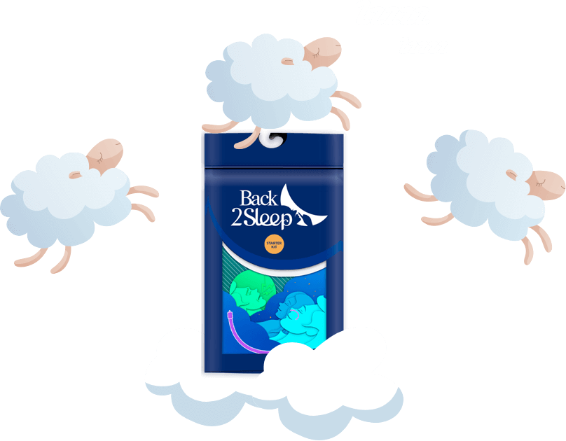
Intranasal Devices
The Back2Sleep intranasal stent maintains nasal airway patency, reducing snoring and mild apnea. Discreet, portable alternative.
Nerve Stimulation
Hypoglossal nerve stimulator (Inspire®) activates tongue muscles during sleep. For moderate-severe OSA patients who can't tolerate CPAP.
Sleep Apnea & Cardiovascular Risk: Why Testing Matters
Untreated obstructive sleep apnea is a recognized independent cardiovascular risk factor. The repeated oxygen desaturations, sleep fragmentation, and sympathetic nervous system activation create a cascade of physiological stress with serious health consequences.
❤️ Cardiovascular Consequences of Untreated OSA
Associated Health Conditions
- Hypertension: OSA contributes to resistant hypertension; CPAP treatment can reduce blood pressure
- Coronary Artery Disease: Increased risk of heart attacks, especially during sleep
- Heart Failure: Bidirectional relationship—OSA worsens heart failure and vice versa
- Atrial Fibrillation: OSA patients have 4x higher risk; treatment improves rhythm control
- Stroke: 2-3x increased risk; nocturnal hypoxemia damages cerebral vasculature
- Type 2 Diabetes: OSA independently increases insulin resistance
- Metabolic Syndrome: Strong association with obesity, dyslipidemia, and glucose intolerance
🩺 The Good News: Treatment Reverses Risk
Research from Johns Hopkins and other institutions demonstrates that consistent CPAP use reduces cardiovascular risk. A 2014 study showed blood pressure reductions in OSA patients using CPAP every night. Early diagnosis through respiratory polygraphy enables timely intervention before irreversible cardiovascular damage occurs.
Frequently Asked Questions
For diagnosing obstructive sleep apnea specifically, respiratory polygraphy demonstrates high accuracy—studies report 93% sensitivity and 100% specificity when proper clinical selection criteria are applied. However, polygraphy may underestimate AHI because it uses recording time rather than actual sleep time. For suspected OSA in patients without significant comorbidities, polygraphy is considered a valid diagnostic alternative to PSG.
Generally, yes—but with important exceptions. Continue prescribed medications unless specifically instructed otherwise by your physician. However, avoid sedatives, sleep aids, and alcohol before the test, as these can artificially worsen sleep apnea by relaxing throat muscles. If you regularly use sedatives for sleep, discuss with your doctor whether to skip them for the test night.
Don't worry—you don't need a "perfect" night's sleep. Most polygraphy devices require only 4-6 hours of recording for adequate analysis. If you experience a particularly poor night (illness, unusual stress, significant insomnia), inform your physician—they may recommend repeating the study. Interestingly, sleep deprivation can sometimes worsen apnea, making the condition more apparent.
Results typically take 1-2 weeks. After device return, technicians download and process the data, then a sleep physician manually scores and interprets the recording. A comprehensive report is generated and sent to your referring physician, who will schedule a follow-up appointment to discuss findings and treatment recommendations.
Borderline results require clinical correlation. If you have significant symptoms (excessive sleepiness, witnessed apneas) or cardiovascular comorbidities (hypertension, atrial fibrillation), treatment may still be recommended. Your physician may also suggest polysomnography for more precise measurement, particularly if polygraphy may have underestimated your AHI.
While home respiratory polygraphy is used in some pediatric settings, polysomnography remains the gold standard for children. Children have different respiratory physiology, and even mild AHI elevations (≥1) can be significant. Studies show polygraphy may underestimate AHI in younger children particularly. If your child needs sleep apnea evaluation, discuss the most appropriate testing method with your pediatrician or pediatric sleep specialist.
Most insurance plans cover respiratory polygraphy when medically indicated for suspected sleep apnea. Home sleep testing is generally less expensive than laboratory polysomnography, which may actually be preferred by insurers. Check with your insurance provider and ensure your physician documents appropriate clinical indications (symptoms, risk factors) for coverage approval.
Conclusion: Taking Control of Your Sleep Health
Nocturnal ventilatory polygraphy represents a significant advancement in sleep medicine accessibility—bringing diagnostic capability for obstructive sleep apnea directly into patients' homes. This ambulatory examination accurately identifies and quantifies sleep-disordered breathing, enabling timely intervention before serious cardiovascular and metabolic complications develop.
🎯 Key Takeaways
- Respiratory polygraphy is a validated home test for diagnosing obstructive sleep apnea
- The Apnea-Hypopnea Index (AHI) classifies severity: mild (5-14), moderate (15-29), severe (≥30)
- AHI ≥ 30 on polygraphy typically confirms diagnosis without need for polysomnography
- Negative results with high clinical suspicion should prompt polysomnography
- Untreated OSA significantly increases cardiovascular risk—early diagnosis saves lives
- Multiple treatment options exist, from CPAP to oral appliances to intranasal devices
If you experience chronic snoring, witnessed breathing pauses, or unexplained daytime fatigue, don't delay—speak with your healthcare provider about sleep testing. Early diagnosis through respiratory polygraphy is the first step toward better sleep, improved health, and enhanced quality of life.
Struggling with Snoring or Sleep Apnea?
The Back2Sleep intranasal device offers a discreet, comfortable solution for snoring and mild sleep apnea. Clinically tested and easy to use, it helps maintain airway patency throughout the night—no mask, no noise, no electricity required.
Try the Back2Sleep Starter KitAvailable at partner pharmacies | Contact us for more information