Polysomnography Explained: The Complete Sleep Study Guide for Accurate Diagnosis
Struggling with unexplained fatigue, loud snoring, or suspected sleep apnea? Polysomnography—the gold-standard overnight sleep study—provides comprehensive diagnosis by recording brain activity, breathing patterns, heart rate, and movements to identify disorders ranging from sleep apnea to narcolepsy, enabling targeted treatment that restores quality sleep.
If you're experiencing chronic fatigue despite adequate sleep time, loud snoring with breathing pauses, or unexplained daytime sleepiness, you may need polysomnography—a comprehensive overnight medical examination that monitors brain waves, eye movements, heart rhythm, breathing patterns, blood oxygen levels, and body movements throughout an entire sleep cycle. This gold-standard diagnostic tool accurately identifies sleep apnea, insomnia, restless legs syndrome, narcolepsy, and other disorders by providing physicians with detailed physiological data that reveals exactly what's disrupting your sleep architecture. While the examination requires an overnight stay at a specialized sleep center with multiple sensors attached to your body, the precise diagnosis it delivers enables targeted treatment—from CPAP therapy to innovative solutions like the Back2Sleep intranasal orthosis that addresses airway obstruction directly.
Understanding what polysomnography involves, why it's necessary, and how results guide treatment empowers you to take the crucial first step toward diagnosing and resolving sleep disorders that may be silently damaging your health.
Polysomnography at a Glance: Essential Information
| Key Aspect | Essential Details |
|---|---|
| What is polysomnography? | Comprehensive overnight medical examination analyzing sleep through continuous recording of brain activity, eye movements, heart rate, breathing, oxygen saturation, and body movements |
| Primary purpose | Detect and characterize sleep apnea, evaluate pre-surgery risks, monitor treatment effectiveness, diagnose other sleep disorders (RLS, narcolepsy, sleepwalking) |
| Exam procedure | Evening arrival at sleep center, sensor placement, overnight recording under surveillance, morning disconnection, specialist analysis |
| Duration | Full night recording (approximately 8 hours) from bedtime through morning wake-up |
| Recorded parameters | EEG (brain waves), EOG (eye movements), EMG (muscle activity), ECG (heart rhythm), respiratory airflow, oxygen saturation, body position |
| Cost range | €120-€400, partially covered by health insurance depending on medical necessity and policy |
| Pain/invasiveness | Non-invasive, painless—involves only external sensor placement, no needles or internal procedures |
| Alternatives | Ventilatory polygraphy (respiratory-focused), actigraphy (movement-based), home sleep apnea testing (limited scope) |
Sleep Disorders by the Numbers
Understanding Polysomnography: The Gold Standard Sleep Diagnostic Tool
Polysomnography represents the most comprehensive and accurate method for diagnosing sleep disorders, providing physicians with detailed physiological data that reveals exactly what occurs during your sleep cycle. Unlike simple screening tools or questionnaires, this multi-parameter examination simultaneously records numerous bodily functions throughout an entire night, creating a complete picture of your sleep architecture and identifying disruptions invisible to conscious awareness.
Comprehensive Data Collection Parameters
Brain Activity (EEG): Electroencephalogram electrodes placed on the scalp record electrical brain wave patterns, identifying different sleep stages (light, deep, REM) and detecting micro-awakenings or abnormal brain activity during sleep.
Eye Movements (EOG): Electro-oculogram sensors near the eyes track eye movement patterns that distinguish REM (rapid eye movement) sleep from non-REM stages, crucial for identifying sleep stage transitions.
Heart Rhythm (ECG): Electrocardiogram electrodes on the chest monitor heart rate variability, detecting arrhythmias, bradycardia, or tachycardia that may occur during respiratory events or other sleep disturbances.
Respiratory Function: Nasal/oral airflow sensors and chest/abdominal effort belts measure breathing patterns, detecting apneas (complete cessation), hypopneas (partial reduction), and respiratory effort during obstructive events.
Muscle Activity (EMG): Electromyogram sensors on chin and legs record muscle tone and movements, identifying conditions like restless legs syndrome, periodic limb movements, or REM behavior disorder.
Oxygen Saturation: Pulse oximetry (fingertip sensor) continuously measures blood oxygen levels, revealing desaturations that indicate severity of respiratory events and their physiological impact.
Body Position: Position sensors track whether you're sleeping on your back, side, or stomach—critical information since many sleep disorders worsen in specific positions.
Snoring Sounds: Audio recording captures snoring intensity and patterns, correlating sound with respiratory events to characterize upper airway obstruction severity.
What Physicians Can Diagnose from Polysomnography Data
The synchronized recording of these multiple parameters enables sleep specialists to:
- Identify sleep stage architecture: Determine if you're achieving adequate deep sleep and REM sleep percentages, or if sleep fragmentation prevents restorative stages
- Detect respiratory abnormalities: Quantify apnea/hypopnea frequency, duration, and associated oxygen desaturations to diagnose OSA severity
- Recognize movement disorders: Document periodic limb movements, restless legs syndrome episodes, or abnormal REM behavior patterns
- Discover cardiac complications: Identify arrhythmias or heart rhythm disturbances correlated with respiratory events
- Assess micro-arousals: Count brief awakenings (lasting just 3-15 seconds) that fragment sleep without your conscious awareness
Conditions Commonly Diagnosed Through Polysomnography
🏥 Sleep Disorders Identified by Polysomnography:
• Obstructive Sleep Apnea (OSA): Repeated upper airway collapse causing breathing cessation and oxygen desaturation
• Central Sleep Apnea: Brain signal disruption causing breathing pauses without physical obstruction
• Chronic Insomnia: Objective documentation of sleep latency, efficiency, and fragmentation patterns
• Restless Legs Syndrome: Involuntary leg movements disrupting sleep initiation and maintenance
• Periodic Limb Movement Disorder: Repetitive limb jerking throughout night causing micro-arousals
• Narcolepsy: Abnormal REM sleep patterns, rapid sleep onset, and sleep/wake boundary disruptions
• REM Behavior Disorder: Acting out dreams due to loss of normal muscle paralysis during REM sleep
• Sleepwalking/Night Terrors: Documentation of parasomnias emerging from deep sleep stages
• Circadian Rhythm Disorders: Sleep-wake timing misalignment with desired schedule
• Sleep-Related Bruxism: Teeth grinding patterns and associated muscle activity during sleep

When Your Doctor Orders Polysomnography: Common Clinical Indications
Sleep specialists prescribe polysomnography when clinical symptoms, screening questionnaires, or preliminary tests suggest underlying sleep-disordered breathing, movement disorders, or other conditions requiring objective physiological documentation for accurate diagnosis and treatment planning.
Suspected Obstructive Sleep Apnea: The Primary Indication
Sleep apnea represents the most common reason physicians order polysomnography. Classic symptoms warranting sleep study evaluation include:
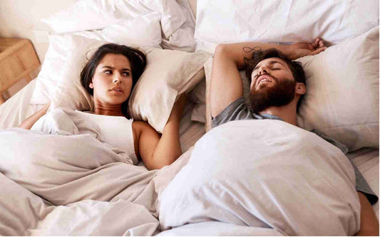
Loud, Chronic Snoring: Particularly snoring audible through closed doors, with gasping or choking sounds indicating airway obstruction
Witnessed Breathing Pauses: Partner reports periods where breathing stops for 10+ seconds followed by sudden gasping or snorting
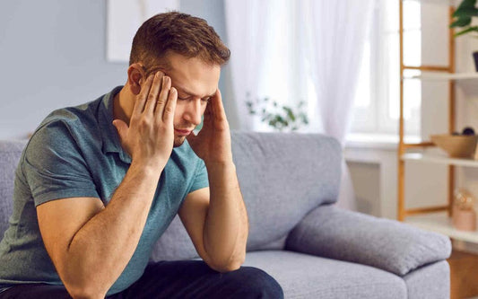
Excessive Daytime Sleepiness: Overwhelming urge to sleep during normal activities despite spending adequate time in bed
Morning Headaches: Waking regularly with headaches across forehead or temples, typically resolving within hours
Polysomnography quantifies apnea severity through the Apnea-Hypopnea Index (AHI)—the number of respiratory events per hour:
- Normal: AHI less than 5 events/hour
- Mild OSA: AHI 5-15 events/hour
- Moderate OSA: AHI 15-30 events/hour
- Severe OSA: AHI greater than 30 events/hour
The study also distinguishes between obstructive apneas (physical airway blockage despite continued respiratory effort) and central apneas (brain signal failure causing absence of breathing effort)—critical information guiding treatment selection.
Pre-Surgical Risk Assessment
Before major surgeries—particularly cardiovascular, neurological, or bariatric procedures—anesthesiologists may order polysomnography to identify undiagnosed sleep apnea that significantly increases surgical complications including:
- Difficult intubation due to anatomical airway narrowing
- Post-operative respiratory depression from anesthesia and pain medications
- Prolonged mechanical ventilation requirements
- Increased cardiovascular events during or after surgery
- Extended hospital stays and ICU admission risks
Identifying OSA pre-operatively allows surgical teams to implement protective strategies including CPAP therapy initiation before surgery, specialized anesthesia protocols, and enhanced post-operative monitoring.
Treatment Effectiveness Monitoring
Patients already receiving treatment for sleep disorders may undergo follow-up polysomnography to verify therapeutic effectiveness and optimize settings:
✓ CPAP Titration Studies: Determine optimal continuous positive airway pressure levels that eliminate apneas without causing discomfort or central apneas from excessive pressure
✓ Oral Appliance Verification: Confirm mandibular advancement devices adequately reduce AHI to acceptable levels
✓ Post-Surgical Assessment: Evaluate success of uvulopalatopharyngoplasty (UPPP) or other airway surgeries in reducing obstruction
✓ Weight Loss Impact: Document improvement in sleep-disordered breathing following significant weight reduction
Diagnosis of Additional Sleep Pathologies
Beyond sleep apnea, polysomnography provides definitive diagnosis for:
Restless Legs Syndrome (RLS) and PLMD: Studies document the frequency and duration of involuntary leg movements during sleep, differentiating between benign movements and those causing significant sleep fragmentation requiring treatment. Learn more about restless legs syndrome.
Narcolepsy: Multiple Sleep Latency Test (MSLT) performed the day following polysomnography measures how quickly you fall asleep during scheduled nap opportunities, identifying pathologically rapid sleep onset and inappropriate REM sleep intrusions characteristic of narcolepsy.
Circadian Rhythm Disorders: Recording reveals delayed or advanced sleep phase patterns where internal biological clock misaligns with desired sleep schedule, causing insomnia at conventional bedtimes. Read about circadian rhythm disorders.
Parasomnias: Video monitoring combined with physiological data captures sleepwalking, sleep terrors, or REM behavior disorder episodes, documenting which sleep stage they emerge from and their frequency—essential for targeted treatment.

Why Choose Back2Sleep After Polysomnography Diagnosis
Diagnosed OSA Solution
If polysomnography confirms mild-to-moderate sleep apnea, Back2Sleep offers effective treatment without bulky CPAP machinery—92% user satisfaction rate.
Simple Implementation
No pressure titration or complex settings required unlike CPAP—insert the device and experience immediate airway support from night one.
Superior Compliance
Comfortable, discreet design encourages nightly use—critical for treatment success compared to 40-50% CPAP abandonment rates due to discomfort.
Cost-Effective Treatment
One-time device purchase versus ongoing CPAP equipment costs, electricity consumption, and replacement supply expenses over years.
The Polysomnography Procedure: What to Expect Step-by-Step
Understanding the examination process helps alleviate anxiety about your upcoming sleep study. While spending a night in an unfamiliar environment with sensors attached may seem uncomfortable, sleep centers are designed to maximize patient comfort and technicians are highly trained to make the experience as natural as possible.
Pre-Study Preparation and Arrival
Arrive at the sleep laboratory in the late afternoon or early evening (typically 7-9 PM), giving technicians time to complete sensor placement before your normal bedtime. Bring:
- Comfortable sleepwear: Two-piece pajamas (not one-piece) to accommodate chest sensors
- Personal toiletries: Toothbrush, face wash, any nighttime hygiene items
- Current medications: Take evening medications as normally prescribed unless physician instructs otherwise
- Sleep diary: If you've been keeping one, bring it to provide context
- Your own pillow: Optional but may help comfort in unfamiliar environment
⚠️ Important Pre-Study Instructions:
• Avoid caffeine after 2 PM on study day—no coffee, tea, energy drinks, or chocolate
• No napping on the day of your study to ensure normal sleep pressure at bedtime
• Skip alcohol for 24 hours before—it alters sleep architecture and can mask or worsen apneas
• Wash hair but don't apply oils, gels, or conditioners that interfere with electrode adhesion
• Continue regular medications unless specifically instructed to hold them
• Remove nail polish from at least one finger for pulse oximeter accuracy
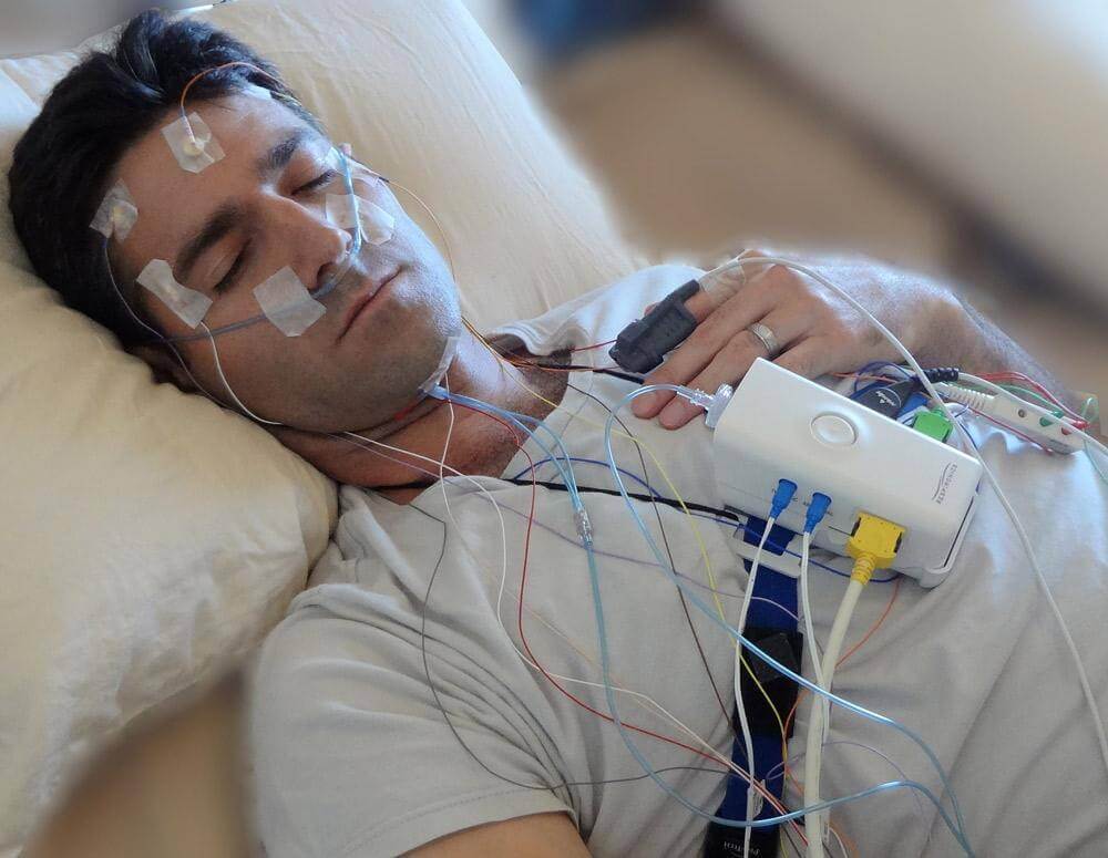
Comprehensive Sensor Placement Process
A registered polysomnographic technologist (RPSGT) will attach numerous sensors using medical-grade adhesive paste and tape. The process takes 30-45 minutes:
Scalp Electrodes (EEG)
6-12 small disc electrodes attached to scalp with conductive paste after mild skin abrasion to reduce impedance. Measures brain wave patterns for sleep staging.
Facial Sensors (EOG/EMG)
Electrodes near outer eye corners track eye movements. Chin sensors monitor muscle tone that decreases during REM sleep.
Chest Monitors (ECG)
3-6 sticky electrodes on chest record heart electrical activity, detecting arrhythmias correlated with respiratory events.
Breathing Sensors
Nasal cannula measures airflow; elastic belts around chest and abdomen detect respiratory effort indicating obstructive versus central apneas.
Leg Electrodes (EMG)
Sensors on both lower legs detect involuntary movements characteristic of restless legs syndrome or periodic limb movement disorder.
Pulse Oximeter
Clip-on finger sensor continuously measures blood oxygen saturation percentage throughout night, revealing desaturations during apneas.
All sensor wires connect to a central junction box near your pillow that amplifies and transmits signals to recording equipment in the control room. While the wires allow movement, you won't have complete freedom to turn as you might at home.
Overnight Recording and Monitoring
Once sensors are placed and tested, you can prepare for bed following your normal nighttime routine. Sleep rooms are designed for comfort with:
- Hotel-quality beds with adjustable firmness options
- Climate control for personal temperature preference
- Complete darkness achieved with blackout curtains or shades
- Sound dampening to minimize external noise disruption
- Infrared cameras for video monitoring without visible light
- Intercom system for communication with technologist if needed
Throughout the night, a technologist monitors your data from an adjacent control room, watching for:
🖥️ Real-Time Monitoring Includes:
• Signal quality verification—adjusting sensors if connections loosen during sleep
• Safety surveillance—responding immediately to any medical emergency
• Event annotation—marking significant findings like apneas, arousals, or position changes
• CPAP titration if ordered—applying graduated pressure increases to determine optimal setting
You can request bathroom breaks anytime via intercom—the technologist will temporarily disconnect you from the recording system.
Morning Disconnection and Departure
Recording typically continues until 6-7 AM or when you naturally awaken. The technologist enters the room, removes all sensors (painless process), and cleanses adhesive residue from your skin with alcohol or remover solution. Most patients depart the sleep center by 7-8 AM.
You can proceed directly to work or daily activities—no recovery time needed. However, some patients report feeling slightly more tired than usual after sleeping in the unfamiliar environment with sensors attached.

Decoding Your Polysomnography Results: Understanding the Report
Following your overnight study, a board-certified sleep medicine physician analyzes hundreds of pages of physiological data to identify patterns, abnormalities, and correlations that reveal your sleep disorder diagnosis. Understanding key metrics helps you comprehend the findings and treatment recommendations.
Sleep Architecture Analysis (EEG Findings)
Brain wave analysis reveals sleep stage distribution throughout the night, comparing your actual percentages to normal adult values:
| Sleep Stage | Normal Percentage | Characteristics | Disruption Implications |
|---|---|---|---|
| Stage 1 (N1) | 2-5% of night | Light sleep, transition from wake, easily disturbed | Excessive N1 indicates fragmented, non-restorative sleep |
| Stage 2 (N2) | 45-55% of night | True sleep onset, memory consolidation begins | Reduced N2 suggests frequent arousals or awakenings |
| Stage 3 (N3/Deep) | 15-25% of night | Slow-wave sleep, physical restoration, growth hormone release | Insufficient deep sleep causes daytime fatigue, impaired healing |
| REM Sleep | 20-25% of night | Rapid eye movements, vivid dreams, emotional processing | REM suppression affects mood, memory, learning capacity |
Sleep efficiency (percentage of time in bed actually sleeping) should exceed 85% in healthy adults. Values below 80% indicate difficulty initiating or maintaining sleep.
Micro-arousals (brief 3-15 second awakenings) are quantified—normal is less than 10 per hour. Excessive arousals fragment sleep without conscious awareness, preventing restorative stages. Learn more about micro-awakenings and their impact.
Respiratory Event Scoring and Severity Grading
The Apnea-Hypopnea Index (AHI) represents the cornerstone metric for sleep-disordered breathing diagnosis:
🫁 Respiratory Event Definitions:
Apnea: Complete cessation of airflow for 10+ seconds with 90%+ flow reduction
Hypopnea: Partial airflow reduction (30-90%) for 10+ seconds causing oxygen desaturation of 3-4% or arousal
RERA: Respiratory effort-related arousal—increased breathing effort not meeting apnea/hypopnea criteria but causing awakening
Respiratory Disturbance Index (RDI): AHI plus RERAs, providing comprehensive upper airway resistance measurement
Event classification matters:
- Obstructive events: Continued or increased respiratory effort against collapsed airway—treated with CPAP, oral appliances, or devices like Back2Sleep
- Central events: Absent respiratory effort due to brain signal failure—requires different treatment approaches like adaptive servo-ventilation
- Mixed events: Begin as central (no effort) then transition to obstructive (effort against closed airway)
Oxygen desaturation patterns reveal physiological impact—brief desaturations to 85-90% differ dramatically from prolonged drops below 80% that stress cardiovascular systems.
Movement and Cardiac Findings
Electromyography (EMG) analysis from leg sensors quantifies:
Periodic Limb Movement Index (PLMI): Number of leg movements per hour. Values exceeding 15/hour with associated arousals indicate Periodic Limb Movement Disorder requiring treatment.
REM muscle tone abnormalities: Normal REM sleep involves near-complete muscle paralysis (atonia). Persistent muscle activity during REM suggests REM Behavior Disorder—a condition where people physically act out dreams, sometimes violently.
Electrocardiography (ECG) reveals:
- Bradycardia (slow heart rate) or tachycardia (fast rate) correlated with respiratory events
- Arrhythmias including atrial fibrillation, premature ventricular contractions, or heart blocks
- Heart rate variability patterns indicating autonomic nervous system dysfunction
The Final Report and Treatment Recommendations
Your comprehensive sleep study report typically arrives within 1-2 weeks, including:
Quantitative Summary: Total sleep time, sleep efficiency, stage percentages, AHI, PLMI, arousal index, minimum oxygen saturation—all key metrics in tabular format
Graphical Hypnogram: Visual timeline showing sleep stage progression, respiratory events, oxygen levels, and position changes throughout the night
Event Examples: Representative waveform samples of apneas, hypopneas, arousals, or limb movements demonstrating characteristic patterns
Clinical Interpretation: Sleep physician's narrative explaining findings, diagnoses, severity grading, and specific treatment recommendations tailored to your results
A follow-up consultation with your sleep specialist reviews results in detail, answers questions, and initiates prescribed therapy—whether CPAP, oral appliance, positional therapy, medications, or innovative solutions like the Back2Sleep intranasal orthosis.
Patient Experiences: Before and After Polysomnography
"I was nervous about sleeping in a lab with all those wires, but the technicians were wonderful. The room was actually more comfortable than I expected. Getting my sleep apnea diagnosis and starting CPAP changed my life—I had no idea how exhausted I'd been for years."
"My polysomnography revealed severe sleep apnea with AHI of 45. I couldn't tolerate CPAP, so my doctor suggested Back2Sleep. It's been eight months and I'm finally sleeping through the night. My wife says I barely snore now."
"The study confirmed I have periodic limb movements causing constant micro-arousals—I was never getting deep sleep. Medication has dramatically improved my sleep quality and daytime energy. I wish I'd done this years ago instead of just accepting fatigue as normal."
"Polysomnography was worth every euro. Turned out I have narcolepsy, not just lazy as everyone thought. Now on proper medication and my career is back on track because I can stay awake during meetings."
Understanding Polysomnography Costs and Insurance Coverage
Complete polysomnography in an accredited sleep laboratory typically costs €120-€400 depending on facility location, study complexity, and whether additional procedures (CPAP titration, MSLT) are included.
Cost Factors Explained
The relatively high price reflects the technical and labor-intensive nature of polysomnography:
- Overnight facility use: Dedicated sleep rooms with specialized equipment, climate control, monitoring systems
- Specialized personnel: Registered polysomnographic technologists (RPSGT) with extensive training monitoring throughout the night
- Advanced equipment: Multi-channel amplifiers, recording systems, video/audio equipment, backup power systems
- Consumable supplies: Electrodes, paste, tape, cleaning materials, disposable sensors for each patient
- Physician interpretation: Board-certified sleep medicine physician spending 1-2 hours analyzing your data
- Accreditation requirements: Facilities must meet strict standards for equipment, personnel qualifications, and quality assurance
Insurance Coverage and Reimbursement
In most European countries, health insurance covers polysomnography when medically necessary—typically requiring:
💶 Insurance Coverage Requirements:
• Physician referral: Sleep study order from general practitioner or specialist documenting medical necessity
• Clinical indications: Symptoms consistent with sleep apnea, narcolepsy, or other disorders warranting diagnostic evaluation
• Pre-authorization: Some insurers require approval before scheduling to confirm coverage eligibility
• Accredited facility: Study must be performed at recognized sleep center meeting national standards
Typical coverage patterns:
- France: Sécurité Sociale covers 70% of approved rate, mutuelle (supplementary insurance) often covers remainder
- Germany: Statutory health insurance (GKV) typically covers full cost for medically indicated studies
- United Kingdom (NHS): Free polysomnography through NHS when referred by physician
- Private insurance: Coverage varies widely—review policy details regarding sleep disorder diagnosis benefits
Out-of-pocket costs depend on your specific insurance policy, with patients typically responsible for copayments (€20-€80) or the balance after primary insurance reimbursement.
Lower-Cost Alternatives for Specific Conditions
Home sleep apnea testing (HSAT) costs €100-€200 and may be appropriate for patients with:
- High pre-test probability of moderate-to-severe OSA without complicating conditions
- No significant cardiovascular, pulmonary, or neurological comorbidities
- Inability to undergo in-laboratory study due to immobility or severe claustrophobia
However, HSAT has limitations: no EEG (can't determine sleep stages), no technologist monitoring, higher failure rates from sensor dislodgement, and inability to diagnose conditions beyond sleep apnea.
Ventilatory polygraphy (respiratory-only monitoring) costs €80-€150 and focuses solely on breathing parameters—appropriate for screening obstructive apnea but missing neurological and movement data that full polysomnography captures.

Alternatives to Full Polysomnography: When Simpler Tests Suffice
While polysomnography remains the diagnostic gold standard, several alternative assessment methods provide useful information for specific situations at lower cost and complexity.
Ventilatory Polygraphy (Cardiorespiratory Sleep Study)
This simplified recording focuses exclusively on breathing during sleep, using 4-7 sensors to measure:
- Nasal and oral airflow (detecting apneas and hypopneas)
- Chest and abdominal respiratory effort (distinguishing obstructive from central events)
- Oxygen saturation via pulse oximetry
- Heart rate from oximeter or ECG lead
- Body position and sometimes snoring sounds
Advantages: Lower cost (€80-€150), can be performed at home, adequate for straightforward OSA screening in patients without complicating medical conditions.
Limitations: No brain wave recording means unable to determine sleep stages, quantify micro-arousals, or diagnose neurological sleep disorders. May underestimate apnea severity if patient doesn't sleep well during monitoring.
Actigraphy: Movement-Based Sleep Assessment
An actigraph resembles a fitness tracker worn on the wrist continuously for 1-2 weeks, using motion sensors to estimate:
- Sleep onset and wake times
- Total sleep duration
- Sleep efficiency (time asleep vs. time in bed)
- Circadian rhythm patterns (sleep-wake timing consistency)
Best for: Evaluating circadian rhythm disorders (delayed/advanced sleep phase), insomnia patterns, treatment response monitoring, shift work sleep disorder assessment.
Limitations: Cannot diagnose sleep apnea, differentiate sleep stages, or detect respiratory/cardiac abnormalities. Estimates sleep based on movement, which may be inaccurate if patient lies still while awake.
Sleep Diary: Subjective Sleep Tracking
A paper or electronic sleep log where patients record daily:
- Bedtime and wake-up time
- Estimated sleep latency (time to fall asleep)
- Number and duration of nighttime awakenings
- Daytime naps
- Medication, caffeine, alcohol use
- Subjective sleep quality rating
Value: Provides context for objective testing, identifies behavioral patterns contributing to insomnia, tracks treatment progress, costs nothing.
Limitations: Completely subjective, prone to recall bias, cannot diagnose physiological disorders requiring objective measurement.
The Back2Sleep Solution: Non-Diagnostic Treatment Approach
For individuals whose polysomnography confirms mild-to-moderate obstructive sleep apnea, or those with primary snoring seeking treatment without formal diagnosis, the Back2Sleep intranasal orthosis offers an evidence-based therapeutic option:
Direct Mechanism: Physically maintains upper airway patency by supporting soft palate—addresses obstruction at the anatomical source
No Pressure Required: Unlike CPAP requiring pressure titration and mask fitting, Back2Sleep works mechanically without air pressure
Superior Compliance: Comfortable, unobtrusive design encourages nightly use—92% user satisfaction versus 40-60% CPAP adherence rates
Cost Advantage: One-time device purchase significantly less expensive than CPAP equipment plus ongoing supply replacements
Frequently Asked Questions About Polysomnography

Take the First Step Toward Diagnostic Clarity
Polysomnography provides the comprehensive physiological data necessary for accurate sleep disorder diagnosis, guiding targeted treatment that addresses your specific condition rather than guessing based on symptoms alone. While the overnight examination requires commitment—sleeping in an unfamiliar environment with multiple sensors—the diagnostic precision it delivers enables evidence-based therapy selection that can transform your sleep quality, daytime functioning, and long-term health outcomes.
If your polysomnography confirms mild-to-moderate obstructive sleep apnea or primary snoring, consider the Back2Sleep intranasal orthosis as an effective, comfortable alternative to CPAP therapy—92% user satisfaction rate and immediate relief from night one without complex equipment or pressure titration.
Explore our comprehensive sleep health library for evidence-based information on all sleep disorders, or contact our sleep specialists for personalized guidance on navigating your polysomnography results and selecting optimal treatment approaches.